Resolving the phylogenetic origin of glioblastoma via multifocal genomic analysis of pre-treatment and treatment-resistant autopsy specimens.

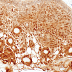
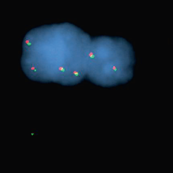
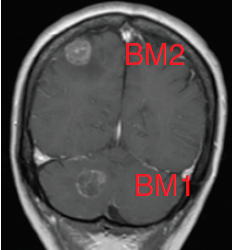
Glioblastomas are malignant neoplasms composed of diverse cell populations. This intratumoral diversity has an underlying architecture, with a hierarchical relationship through clonal evolution from a common ancestor. Therapies are limited by emergence of resistant subclones from this phylogenetic reservoir. To characterize this clonal ancestral origin of recurrent tumors, we determined phylogenetic relationships using whole exome sequencing of pre-treatment IDH1/2 wild-type glioblastoma specimens, matched to post-treatment autopsy samples (n = 9) and metastatic extracranial post-treatment autopsy samples (n = 3). We identified “truncal” genetic events common to the evolutionary ancestry of the initial specimen and later recurrences, thereby inferring the identity of the precursor cell population. Mutations were identified in a subset of cases in known glioblastoma genes such as NF1(n = 3), TP53(n = 4) and EGFR(n = 5). However, by phylogenetic analysis, there were no protein-coding mutations as recurrent truncal events across the majority of cases. In contrast, whole copy-loss of chromosome 10 (12 of 12 cases), copy-loss of chromosome 9p21 (11 of 12 cases) and copy-gain in chromosome 7 (10 of 12 cases) were identified as shared events in the majority of cases. Strikingly, mutations in the TERT promoter were also identified as shared events in all evaluated pairs (9 of 9). Thus, we define four truncal non-coding genomic alterations that represent early genomic events in gliomagenesis, that identify the persistent cellular reservoir from which glioblastoma recurrences emerge. Therapies to target these key early genomic events are needed. These findings offer an evolutionary explanation for why precision therapies that target protein-coding mutations lack efficacy in GBM.

Brastianos PK, Nayyar N, Rosebrock D, Leshchiner I, Gill CM, Livitz D, Bertalan MS, D’Andrea M, Hoang K, Aquilanti E, Chukwueke UN, Kaneb A, Chi A, Plotkin S, Gerstner ER, Frosch MP, Suva ML, Cahill DP, Getz G, Batchelor TT.
NPJ Precis Oncol. 2017 Sep 18;1(1):33. doi: 10.1038/s41698-017-0035-9. eCollection 2017.

DMD genomic deletions characterize a subset of progressive/higher-grade meningiomas with poor outcome.

Progressive meningiomas that have failed surgery and radiation have a poor prognosis and no standard therapy. While meningiomas are more common in females overall, progressive meningiomas are enriched in males. We performed a comprehensive molecular characterization of 169 meningiomas from 53 patients with progressive/high-grade tumors, including matched primary and recurrent samples. Exome sequencing in an initial cohort (n = 24) detected frequent alterations in genes residing on the X chromosome, with somatic intragenic deletions of the dystrophin-encoding and muscular dystrophy-associated DMD gene as the most common alteration (n = 5, 20.8%), along with alterations of other known X-linked cancer-related genes KDM6A (n =2, 8.3%), DDX3X, RBM10 and STAG2 (n = 1, 4.1% each). DMD inactivation (by genomic deletion or loss of protein expression) was ultimately detected in 17/53 progressive meningioma patients (32%). Importantly, patients with tumors harboring DMD inactivation had a shorter overall survival (OS) than their wild-type counterparts [5.1 years (95% CI 1.3-9.0) vs. median not reached (95% CI 2.9-not reached, p = 0.006)]. Given the known poor prognostic association of TERT alterations in these tumors, we also assessed for these events, and found seven patients with TERT promoter mutations and three with TERT rearrangements in this cohort (n = 10, 18.8%), including a recurrent novel RETREG1-TERT rearrangement that was present in two patients. In a multivariate model, DMD inactivation (p = 0.033, HR = 2.6, 95% CI 1.0-6.6) and TERT alterations (p = 0.005, HR = 3.8, 95% CI 1.5-9.9) were mutually independent in predicting unfavorable outcomes. Thus, DMD alterations identify a subset of progressive/high-grade meningiomas with worse outcomes.

Juratli TA1,2,3, McCabe D4,5, Nayyar N2, Williams EA1,6, Silverman IM7, Tummala SS1, Fink AL1, Baig A6, Martinez-Lage M6, Selig MK6, Bihun IV2, Shankar GM1, Penson T1, Lastrapes M4,5, Daubner D8, Meinhardt M9, Hennig S3, Kaplan AB2, Fujio S1, Kuter BM2, Bertalan MS2, Miller JJ1, Batten JM6, Ely HA7, Christiansen J7, Baretton GB9, Stemmer-Rachamimov AO6, Santagata S10, Rivera MN6, Barker FG 2nd1, Schackert G3, Wakimoto H1, Iafrate AJ6, Carter SL4,5, Cahill DP1, Brastianos PK11.
Acta Neuropathol. 2018 Nov;136(5):779-792. doi: 10.1007/s00401-018-1899-7. Epub 2018 Aug 19.

MYD88 L265P mutation and CDKN2A loss are early mutational events in primary central nervous system diffuse large B-cell lymphomas.

The genetic alterations that define primary central nervous system lymphoma (PCNSL) are incompletely elucidated, and the genomic evolution from diagnosis to relapse is poorly understood. We performed whole-exome sequencing (WES) on 36 PCNSL patients and targeted MYD88 sequencing on a validation cohort of 27 PCNSL patients. We also performed WES and phylogenetic analysis of 3 matched newly diagnosed and relapsed tumor specimens and 1 synchronous intracranial and extracranial relapse. Immunohistochemistry (IHC) for programmed death-1 ligand (PD-L1) was performed on 43 patient specimens. Combined WES and targeted sequencing identified MYD88 mutation in 67% (42 of 63) of patients, CDKN2Abiallelic loss in 44% (16 of 36), and CD79b mutation in 61% (22 of 36). Copy-number analysis demonstrated frequent regions of copy loss (ie, CDKN2A), with few areas of amplification. CD79b mutations were associated with improved progression-free and overall survival. We did not identify amplification at the PD-1/PD-L1 loci. IHC for PD-L1 revealed membranous expression in 30% (13 of 43) of specimens. Phylogenetic analysis of paired primary and relapsed specimens identified MYD88mutation and CDKN2A loss as early clonal events. PCNSL is characterized by frequent mutations within the B-cell receptor and NF-κB pathways. The lack of PD-L1 amplifications, along with membranous PD-L1 expression in 30% of our cohort, suggests that PD-1/PD-L1 inhibitors may be useful in a subset of PCNSL. WES of PCNSL provides insight into the genomic landscape and evolution of this rare lymphoma subtype and potentially informs more rational treatment decisions.

Nayyar N1,2, White MD3,4, Gill CM1, Lastrapes M1,2,5, Bertalan M1, Kaplan A1, D’Andrea MR1, Bihun I1, Kaneb A1, Dietrich J1,3,4, Ferry JA6, Martinez-Lage M3,6, Giobbie-Hurder A5, Borger DR1,3, Rodriguez FJ7, Frosch MP3,6, Batchelor E1, Hoang K1, Kuter B1, Fortin S1, Holdhoff M7, Cahill DP1,3,4,8, Carter S2,5,9, Brastianos PK1,2,3,4, Batchelor TT1,3,4.
Blood Adv. 2019 Feb 12;3(3):375-383. doi: 10.1182/bloodadvances.2018027672.

Upfront Surgical Resection of Melanoma Brain Metastases Provides a Bridge Toward Immunotherapy-Mediated Systemic Control.

Background.

Immune checkpoint blockade has systemic efficacy in patients with metastatic melanoma, including those with brain metastases (MBMs). However, immunotherapy‐induced intracranial tumoral inflammation can lead to neurologic compromise, requiring steroids, which abrogate the systemic efficacy of this approach. We investigated whether upfront neurosurgical resection of MBM is associated with a therapeutic advantage when performed prior to initiation of immunotherapy.

Material and Methods.

An institutional review board‐approved, retrospective study identified 142 patients with MBM treated with immune checkpoint blockade between 2010 and 2016 at Massachusetts General Hospital, of whom 79 received surgery. Patients were classified based on the temporal relationship between immunotherapy, surgery, and development of central nervous system metastases. Overall survival (OS) was calculated from the date of diagnosis of MBM until death from any cause. Multivariate model building included a prognostic Cox model of OS, the effect of immunotherapy and surgical sequencing on OS, and the effect of immunotherapy and radiation sequencing on OS.

Results.

The 2‐year overall survival for patients treated with cytotoxic T‐lymphocyte antigen 4, programmed death 1, or combinatorial blockade was 19%, 54%, and 57%, respectively. Among immunotherapy‐naïve melanoma brain metastases, surgery followed by immunotherapy had a median survival of 22.7 months (95% confidence interval [CI], 12.6–39.2) compared with 10.8 months for patients treated with immunotherapy alone (95% CI, 7.8–16.3) and 9.4 months for patients treated with immunotherapy followed by surgery (95% CI, 4.1 to ∞; p = .12). On multivariate analysis, immunotherapy‐naïve brain metastases treated with immunotherapy alone were associated with increased risk of death (hazard ratio, 1.72; 95% CI, 1.00–2.99) compared with immunotherapy‐naïve brain metastases treated with surgery followed by immunotherapy.

Conclusion.

In treatment‐naïve patients, early surgical resection for local control should be considered prior to commencing immunotherapy. A prospective, randomized trial comparing the sequence of surgery and immunotherapy for treatment‐naïve melanoma brain metastases is warranted.

Alvarez-Breckenridge C, Giobbie-Hurder A, Gill CM, Bertalan M, Stocking J, Kaplan A, Nayyar N, Lawrence DP, Flaherty KT, Shih HA, Oh K, Batchelor TT, Cahill DP, Sullivan R, Brastianos P.
Oncologist. 2019 Feb 22. pii: theoncologist.2018-0306. doi: 10.1634/theoncologist.2018-0306. [Epub ahead of print]

The dual PI3K/mTOR-pathway inhibitor GDC-0084 achieves antitumor activity in PIK3CA-mutant breast cancer brain metastases.

Purpose: Previous studies have shown that the PI3K/Akt/mTOR-pathway is activated in up to 70% of breast cancer brain metastases, but there are no approved agents for affected patients. GDC-0084 is a brain penetrant, dual PI3K/mTOR-inhibitor that has shown promising activity in a preclinical model of glioblastoma. The aim of this study was to analyze the efficacy of PI3K/mTOR blockade in breast cancer brain metastases models. Experimental Design: The efficacy of GDC-0084 was evaluated in PIK3CA-mutant and PIK3CA-wildtype breast cancer cell lines and the isogenic pairs of PIK3CA-wildtype and -mutant (H1047R/+) MCF10A cells in vitro. In vitro studies included cell viability and apoptosis assays, cell cycle analysis and Western blots. In vivo, the effect of GDC-0084 was investigated in breast cancer brain metastasis xenograft mouse models and assessed by bioluminescent imaging and immunohistochemistry. Results: In vitro, GDC-0084 considerably decreased cell viability, induced apoptosis and inhibited phosphorylation of Akt and p70 S6 kinase in a dose-dependent manner in PIK3CA-mutant breast cancer brain metastatic cell lines. In contrast, GDC-0084 led only to growth inhibition in PIK3CA-wildtype cell lines in vitro. In vivo, treatment with GDC-0084 markedly inhibited the growth of PIK3CA-mutant, with accompanying signaling changes, and not PIK3CA-wildtype brain tumors. Conclusions: The results of this study suggest that the brain-penetrant PI3K/mTOR-targeting GDC-0084 is a promising treatment option for breast cancer brain metastases with dysregulated PI3K/mTOR signaling pathway conferred by activating PIK3CA mutations. A national clinical trial is planned to further investigate the role of this compound in patients with brain metastases.

Ippen FM, Alvarez-Breckenridge CA, Kuter BM, Fink AL, Bihun IV, Lastrapes M, Penson T, Schmidt SP, Wojtkiewicz GR, Ning J, Subramanian M, Giobbie-Hurder A, Martinez-Lage M, Carter SL, Cahill DP, Wakimoto H, Brastianos PK.
Clin Cancer Res. 2019 Feb 22. pii: clincanres.3049.2018. doi: 10.1158/1078-0432.CCR-18-3049. [Epub ahead of print]

Targeted sequencing of SMO and AKT1 in anterior skull base meningiomas (2016)

Meningiomas located in the skull base are surgically challenging. Recent genomic research has identified oncogenic SMOand AKT1 mutations in a small subset of meningiomas. METHODS The authors performed targeted sequencing in a large cohort of patients with anterior skull base meningiomas (n = 62) to better define the frequency of SMO and AKT1mutations in these tumors. RESULTS The authors found SMO mutations in 7 of 62 (11%) and AKT1 mutations in 12 of 62 (19%) of their cohort. Of the 7 meningiomas with SMO mutations, 6 (86%) occurred in the olfactory groove. Meningiomas with an SMO mutation presented with significantly larger tumor volume (70.6 ± 36.3 cm3) compared with AKT1-mutated (18.2 ± 26.8 cm3) and wild-type (22.7 ± 23.9 cm3) meningiomas, respectively. CONCLUSIONS Combined, these data demonstrate clinically actionable mutations in 30% of anterior skullbasemeningiomas and suggest an association between SMO mutation status and tumor volume. Genotyping of SMO and AKT1 is likely to be high yield in anterior skull base meningiomas with available surgical tissue.

Clinical and radiographic response following targeting of BCAN-NTRK1 fusion in glioneuronal tumor (2017)

Glioneuronal tumors constitute a histologically diverse group of primary central nervous system neoplasms that are typically slowgrowing and managed conservatively. Genetic alterations associated with glioneuronal tumors include BRAF mutations and oncogenic fusions. To further characterize this group of tumors, we collected a cohort of 26 glioneuronal tumors and performed indepth genomic analysis. We identified mutations in BRAF (34%) and oncogenic fusions (30%), consistent with previously published reports. In addition, we discovered novel oncogenic fusions involving members of the NTRK gene family in a subset of our cohort. One-patient with BCAN exon 13 fused to NTRK1 exon 11 initially underwent a subtotal resection for a 4th ventricular glioneuronal tumor but ultimately required additional therapy due to progressive, symptomatic disease. Given the patient’s targetable fusion, the patient was enrolled on a clinical trial with entrectinib, a pan-Trk, ROS1, and ALK (anaplastic lymphoma kinase) inhibitor. The patient was treated for 11 months and during this time volumetric analysis of the lesion demonstrated a maximum reduction of 60% in the contrast-enhancing tumor compared to his pre-treatment magnetic resonance imaging study. The radiologic response was associated with resolution of his clinical symptoms and was maintained for 11 months on treatment. This report of a BCAN-NTRK1 fusion in glioneuronal tumors highlights its clinical importance as a novel, targetable alteration.

Diagnosis and management of craniopharyngiomas in the era of genomics and targeted therapy (2016)

Craniopharyngiomas are rare intracranial neoplasms that pose clinical challenges due to their location adjacent to vital structures. The authors have previously shown high mutation rates of BRAF V600E in papillary craniopharyngioma and of CTNNB1 in adamantinomatous craniopharyngioma. These activating driver mutations are potential therapeutic targets, and the authors have recently reported a significant response to BRAF/MEK inhibition in a patient with multiply recurrent PCP. As these targetable mutations warrant prospective research, the authors will be conducting a national National Cancer Institute-sponsored multicenter clinical trial to investigate BRAF/MEK inhibition in the treatment of craniopharyngioma. In this new eraof genomic discovery, the treatment paradigm of craniopharyngioma is likely to change.

Alterations in Pericyte Subpopulations Are Associated with Elevated Blood-Tumor Barrier Permeability in Experimental Brain Metastasis of Breast Cancer (2016)

The blood-brain barrier (BBB) is modified to a blood-tumor barrier (BTB) as a brain metastasisdevelops from breast or other cancers. We (i) quantified the permeability of experimental brainmetastases, (ii) determined the composition of the BTB, and (iii) identified which elements of the BTB distinguished metastases of lower permeability from those with higher permeability.

Management of Brain Metastases in Patients With Melanoma (2016)

Melanoma is the third most common systemic cancer that leads to brain metastases. The annual incidence of melanoma has increased over time, with brain metastases developing in 40% to 50% of patients with advanced melanoma. Traditional management of melanoma-related brain metastases has focused on symptom control as a result of the significant neurologic morbidity associated with the disease. Median overall survival for these patients, if untreated, is approximately 3 months. As with other brain metastases, a multidisciplinary treatment approach that includes surgery and radiation therapy is typically used, with historically little role for systemic, cytotoxic therapy. During the past decade, advancement within the field of genomics has led to the identification of melanoma-specific mutations, namely, v-Raf murine sarcoma viral oncogene homolog B and neuroblastoma RAS viral oncogene homolog, as well as to the development of agents that target these driver mutations. In addition, the advent of immunotherapies, specifically, agents that target cytotoxic T-lymphocyte antigen-4, anti–programmed death-1, and programmed death ligand-1, has increased the potential therapeutic options available to patients with both systemic and brain disease. With these advances, early trials have demonstrated improved overall survival in patients with brain metastases who receive these therapies either as single agents or as part of multimodality treatment regimens.